


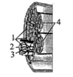
The given figure shows the diagrammatic cross sectional view of a muscle with their parts marked as 1, 2, 3 and 4. Which part is held together by a common collagenous connective tissue layer?
A. 1
B. 2
C. 3
D. 4
Answer
388.8k+ views
Hint: The complete figure given represents a section of muscle anatomy. It depicts the constitution of a skeletal or a voluntary muscle. Along with it, it shows the intervention of the blood vessels that provide nutrients to the muscle.
Complete Step by Step Answer:
Figure 1 depicts muscle fibre.
Muscle fibres are simply another term for muscle cells surrounded by a connective tissue layer called endomysium. These are made up of myofibrils. Myofibrils, in turn, are composed of actin and myosin filaments. A bundle of muscle fibres constitutes a fascicle which is wrapped around by a connective layer called perimysium.
Figure 2 depicts sarcolemma. It refers to the plasma membrane of a muscle fibre. This plays a significant role in contraction of voluntary muscles. Other than that, it performs a basic function as that of a plasma membrane, that is, providing a barrier to the cell from the external environment.
Figure 3 depicts blood capillary. The vascular system in a skeletal muscle is complex. Each muscle cell /fibre is provided with a capillary .
Figure 4 depicts fascicles. As stated above. It is a bundle of muscle fibres. The connective tissue/perimysium that it is surrounded with is collagenous.
Thus, the correct answer is D: 4
Note: Connective tissue, as the name implies, are the tissues to connect tissues, of same or different origin. There are 4 major types of connective tissues: Loose connective tissue, Dense connective tissue, Specialised connective tissue.
Recently Updated Pages
Master Class 12 Economics: Engaging Questions & Answers for Success
Master Class 12 English: Engaging Questions & Answers for Success
Master Class 12 Social Science: Engaging Questions & Answers for Success
Master Class 12 Maths: Engaging Questions & Answers for Success
Master Class 12 Physics: Engaging Questions & Answers for Success
Master Class 9 General Knowledge: Engaging Questions & Answers for Success
Trending doubts
According to phylogenetic classification systems organisms class 11 biology NEET_UG
The common characteristics between tomato and potato class 11 biology NEET_UG
Which are the Top 10 Largest Countries of the World?
One Metric ton is equal to kg A 10000 B 1000 C 100 class 11 physics CBSE
Explain the Treaty of Vienna of 1815 class 10 social science CBSE
Draw a labelled sketch of the human eye class 12 physics CBSE