


Long Answer Question: Describe different parts of the human eye.
Answer
582.6k+ views
Hint: In humans, the human eye is a specialised sense organ capable of obtaining sensory images that are then transported to the brain . The human eye is an organ that responds to light and enables perception of light, vision of colour and perception of depth.
Complete Answer:
In the skull, the eyes rest in cone-shaped cavities called sockets that are surrounded by 6 muscles that control motion and several layers of fatty tissue that help protect and provide flexibility to the eye. Often contributing to this initiative are eyebrows, eyelashes and eyelids.
The eye itself consists of 10 general components that all work together every day to keep us seeing well.
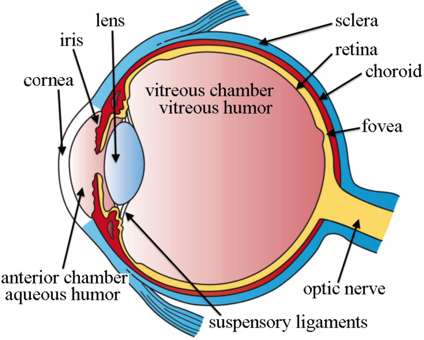
Sclera: The white of the eye is the Sclera. The exterior is white and smooth.
- Flexibility adds power
- Continuous with an optic nerve sheath.
- Attached to it by tendons.
The Cornea: The obvious bulging surface in front of the eye is the cornea. It is the eye 's principal refractive surface.
- Primary eye refractive surface.
- Refraction index: n = 1.37
- Normally transparent and evenly dense.
- Almost in avascular form.
- Richly supplied with fibres of nerves.
- Sensitive to foreign bodies, cold air, irritation of chemicals.
- Aqueous humour and nutrition.
- Oxygen exchange and water content are maintained by Tears.
- Tears prevent dispersion and improve the quality of optics.
Anterior & Posterior Chambers: Chamber between the cornea and the iris is anterior,
- Chamber between the iris and the lens is posterior,
- Refraction index: n = 1.33
- Specific aqueous viscosity of just above 1.0 (like water, hence the name).
- The 15-18 mm mercury pressure maintains the shape of the eye and the spacing of the elements.
- Aqueous humour is created from plasma blood.
- It takes approximately an hour to renew.
- Glaucoma is caused by increased fluid pressure in the eye due to a reduction or blockage in the anterior to posterior aqueous chambers of the eye.
Pupil / Iris: Iris is extensively pigmented. To constrict or dilate the pupil, the Sphincter muscle.
- The pupil is the hole where light passes through.
- The diameter of the pupil ranges from about 3-7 mm.
- The area 7-38 square mm.
- Eye colour (brown, green , blue, etc.) depends on the amount of melanin pigment and its distribution.
Lens: Transparent body with elastic capsule enclosure, composed of Proteins and water.
Consists of layers with a firm nucleus, such as an onion, soft cortex.
- Refractive index for the gradient (1.38-1.40).
- Via ciliary muscles, young people can change the shape of the lens.
- Muscle contraction causes lenses to bulge.
- The lens can no longer change shape at around age 50.
- With age, it becomes more yellow: Cataracts.
Vitreous Humor: The space between the retina and the lens is filled by vitreous humor.
- Gelatinous transparent body.
- Specific viscosity of 1.8-2.0 (consistency similar to jelly)
- Refraction index, n = 1.33
- Nutrition from retinal vessels, aqueous body, ciliary body.
- Floaters, in the vitreous shadows of sloughed off material / debris.
- Keeps the shape of the eye.
Retina: It is a layer that is light-sensitive and is made up of numerous nerve cells. It transforms the lens-formed images into electrical impulses. Through optic nerves, these electrical impulses are then conveyed to the brain.
Optic nerves: There are two types of optic nerves. Cones and rods include these.
Cones: The nerve cells that are more susceptible to bright light are cones. In detailed central and colour vision, they assist.
Rods: The optic nerve cells that are more vulnerable to dim lights are rods. They contribute to peripheral vision.
There are no sensory nerve cells at the junction of the optic nerve and the retina. At that point, no vision is possible, and it is known as a blind spot.
The Fovea: Fovea is the place of the central gaze on the retina. The fovea is the retinal locus of this central fixation when you look directly, or fixate, at a stimulus.
In Human Fovea, there are only cones (no rods). They are thinner, elongated and packed very tightly. Therefore, the fovea is the location of highest visual acuity and best vision of colour.
The Macula: A pigment called the macula is the covering of the fovea. It is believed that the macula acts over the fovea as a protective philtre that absorbs blue and ultraviolet radiation. This pigment varies from observer to superior and is a source of individual colour vision variation. We usually do not notice the macula filtering, but we can notice its presence causing what is known as Maxwell's spot under special conditions.
Note: The human eye, much like the electronic device, also focuses and allows images to be produced in light. So basically, after passing through different mediums like the cornea, crystalline lens, aqueous humour, the lens, and vitreous humour, light rays that are deflected from or by distant objects land on the retina.
Complete Answer:
In the skull, the eyes rest in cone-shaped cavities called sockets that are surrounded by 6 muscles that control motion and several layers of fatty tissue that help protect and provide flexibility to the eye. Often contributing to this initiative are eyebrows, eyelashes and eyelids.
The eye itself consists of 10 general components that all work together every day to keep us seeing well.
Sclera: The white of the eye is the Sclera. The exterior is white and smooth.
- Flexibility adds power
- Continuous with an optic nerve sheath.
- Attached to it by tendons.
The Cornea: The obvious bulging surface in front of the eye is the cornea. It is the eye 's principal refractive surface.
- Primary eye refractive surface.
- Refraction index: n = 1.37
- Normally transparent and evenly dense.
- Almost in avascular form.
- Richly supplied with fibres of nerves.
- Sensitive to foreign bodies, cold air, irritation of chemicals.
- Aqueous humour and nutrition.
- Oxygen exchange and water content are maintained by Tears.
- Tears prevent dispersion and improve the quality of optics.
Anterior & Posterior Chambers: Chamber between the cornea and the iris is anterior,
- Chamber between the iris and the lens is posterior,
- Refraction index: n = 1.33
- Specific aqueous viscosity of just above 1.0 (like water, hence the name).
- The 15-18 mm mercury pressure maintains the shape of the eye and the spacing of the elements.
- Aqueous humour is created from plasma blood.
- It takes approximately an hour to renew.
- Glaucoma is caused by increased fluid pressure in the eye due to a reduction or blockage in the anterior to posterior aqueous chambers of the eye.
Pupil / Iris: Iris is extensively pigmented. To constrict or dilate the pupil, the Sphincter muscle.
- The pupil is the hole where light passes through.
- The diameter of the pupil ranges from about 3-7 mm.
- The area 7-38 square mm.
- Eye colour (brown, green , blue, etc.) depends on the amount of melanin pigment and its distribution.
Lens: Transparent body with elastic capsule enclosure, composed of Proteins and water.
Consists of layers with a firm nucleus, such as an onion, soft cortex.
- Refractive index for the gradient (1.38-1.40).
- Via ciliary muscles, young people can change the shape of the lens.
- Muscle contraction causes lenses to bulge.
- The lens can no longer change shape at around age 50.
- With age, it becomes more yellow: Cataracts.
Vitreous Humor: The space between the retina and the lens is filled by vitreous humor.
- Gelatinous transparent body.
- Specific viscosity of 1.8-2.0 (consistency similar to jelly)
- Refraction index, n = 1.33
- Nutrition from retinal vessels, aqueous body, ciliary body.
- Floaters, in the vitreous shadows of sloughed off material / debris.
- Keeps the shape of the eye.
Retina: It is a layer that is light-sensitive and is made up of numerous nerve cells. It transforms the lens-formed images into electrical impulses. Through optic nerves, these electrical impulses are then conveyed to the brain.
Optic nerves: There are two types of optic nerves. Cones and rods include these.
Cones: The nerve cells that are more susceptible to bright light are cones. In detailed central and colour vision, they assist.
Rods: The optic nerve cells that are more vulnerable to dim lights are rods. They contribute to peripheral vision.
There are no sensory nerve cells at the junction of the optic nerve and the retina. At that point, no vision is possible, and it is known as a blind spot.
The Fovea: Fovea is the place of the central gaze on the retina. The fovea is the retinal locus of this central fixation when you look directly, or fixate, at a stimulus.
In Human Fovea, there are only cones (no rods). They are thinner, elongated and packed very tightly. Therefore, the fovea is the location of highest visual acuity and best vision of colour.
The Macula: A pigment called the macula is the covering of the fovea. It is believed that the macula acts over the fovea as a protective philtre that absorbs blue and ultraviolet radiation. This pigment varies from observer to superior and is a source of individual colour vision variation. We usually do not notice the macula filtering, but we can notice its presence causing what is known as Maxwell's spot under special conditions.
Note: The human eye, much like the electronic device, also focuses and allows images to be produced in light. So basically, after passing through different mediums like the cornea, crystalline lens, aqueous humour, the lens, and vitreous humour, light rays that are deflected from or by distant objects land on the retina.
Recently Updated Pages
Master Class 11 Social Science: Engaging Questions & Answers for Success
Master Class 11 Physics: Engaging Questions & Answers for Success
Master Class 11 Maths: Engaging Questions & Answers for Success
Master Class 11 Economics: Engaging Questions & Answers for Success
Master Class 11 Computer Science: Engaging Questions & Answers for Success
Master Class 11 Chemistry: Engaging Questions & Answers for Success
Trending doubts
One Metric ton is equal to kg A 10000 B 1000 C 100 class 11 physics CBSE
How many bones are in the spine class 11 biology CBSE
1 Quintal is equal to a 110 kg b 10 kg c 100kg d 1000 class 11 physics CBSE
There are 720 permutations of the digits 1 2 3 4 5 class 11 maths CBSE
State and prove Bernoullis theorem class 11 physics CBSE
Difference Between Prokaryotic Cells and Eukaryotic Cells