


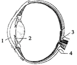
In the given figure of the eye, a few parts are labelled as 1, 2, 3 and 4. Select the option which shows the correct identification of the part with its characteristics.
A) 1: Choroid, contains ganglion cells, bipolar cells and photoreceptor cells.
B) 2: Iris, is responsible for controlling the diameter and size of the pupil and thus the amount of light reaching the retina.
C) 3: Blind spot, is a yellowish-pigmented spot called the macula lutea with a central pit called the fovea.
D) 4: Cornea, is a transparent front part of the eye that covers the iris, pupil, and anterior chamber.
Answer
356.7k+ views
Hint:
The eyes are complex sense organs. Each eye has a layer of receptors and a system of nerves that conducts impulses from the receptors to the brain. The eyes are spherical structures that are filled with fluid.
Complete step by step answer:
The figure represents a human eye and part (1) represents the cornea, part (2) represents the iris, part (3) represents a blind spot and part (4) represents the choroid. The transparent coat that covers the iris is named the cornea. It mediates the focusing of light on the retina as it is curved in shape. The middle coat of the cornea consists of collagen fibres and fibroblasts, and the inner surface is the simple squamous epithelium.
The iris is the pigmented and coloured portion of the eye. The round opening in the centre of the iris through which light enters the interior portions of the eye is a pupil. The iris contains circular muscles that constrict and radial muscles dilate the pupil. The choroid provides nutrients as well as oxygen to the structures in the eye as it is a vascular layer. Blind spot: An inner layer of ganglion cells is present. The point on the retina at which the optic nerve leaves and through which blood vessels pass is the optic disc. This region is called a blind spot. This area has no rods and cones so no image can be detected.
Option ‘B’ is correct
Note:
A small depression in the centre of the macula lutea (yellowish pigmented spot) is called fovea centralis. In it, the cones are packed densely. There are no blood vessels. Consequently, the fovea is the point where visual acuity is the greatest.
Recently Updated Pages
Master Class 9 General Knowledge: Engaging Questions & Answers for Success
Master Class 9 Social Science: Engaging Questions & Answers for Success
Master Class 9 English: Engaging Questions & Answers for Success
Master Class 9 Maths: Engaging Questions & Answers for Success
Master Class 9 Science: Engaging Questions & Answers for Success
Class 9 Question and Answer - Your Ultimate Solutions Guide
Trending doubts
Which are the Top 10 Largest Countries of the World?
What is BLO What is the full form of BLO class 8 social science CBSE
The value of 6 more than 7 is A 1 B 1 C 13 D 13 class 7 maths CBSE
One Metric ton is equal to kg A 10000 B 1000 C 100 class 11 physics CBSE
There are 720 permutations of the digits 1 2 3 4 5 class 11 maths CBSE
What is the median of the first 10 natural numbers class 10 maths CBSE