


Human eye lens is
A. Spherical and can be moved forward
B. Biconvex and cannot be moved forward
C. Spherical and cannot be moved forward
D. Biconvex and can be moved forward
Answer
635.1k+ views
Hint: The eye lens shape is similar to that of lenses used in microscopes. The lens is able to change its aperture depending upon the position of the object with the help of ciliary muscles. No movement is necessary.
Complete answer:
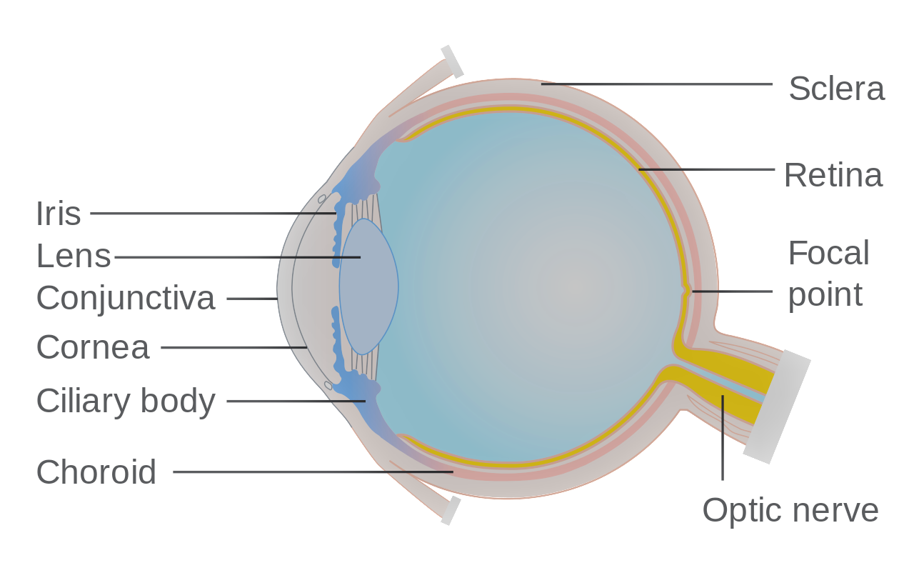
The human eye lens is a fibrous crystalline structure formed of alpha and beta crystalline proteins. It is colorless, transparent, and enclosed in a lens membrane. It is biconvex and cannot be moved forward because it is held back by the suspensory ligaments.
Additional Information:
Eye lens:
- The suspensory ligaments are also known as Zonula of Zinn. In humans the eye lens is biconvex but it is elliptical or subspherical in case of frogs.
- The eyeball is divided into two chambers by the lens, the external chamber is filled with aqueous humor which is watery and secreted by the ciliary body. The internal chamber is also known as the vitreous chamber filled with vitreous jelly which is also known as Wharton’s jelly, besides water it contains mucoprotein and hyaluronic acid.
Protective parts of the eye are:
- Eyebrows: Hair like structures directed outward, they carry away sweat and raindrops trickling from above forehead away from the eye.
- Eyelids or Palpebrae: Two eyelids are present in humans but on the upper eyelid can be moved.
- Eyelashes: These are stiff hair-like structures present at the free edge of eyelids. Help to prevent entry or falling of dust particles, and small organisms into the eyes.
- Eye glands: Several secretions help to keep the eye surface moist and secretions like lysozyme act as an antibacterial agent.
So, the correct answer is,” Human eye lens is biconvex and cannot be moved forward”
Note:
The eyeball has attachments to six extrinsic muscles which include anterior rectus, posterior rectus, inferior rectus, superior rectus, inferior oblique muscles, and inferior oblique muscles. In frogs, out of the two eyelids, the lower eyelid is movable, unlike humans. The nictitating membrane which is a vestigial organ in humans is still functional in frogs and birds. It helps to protect the eye. The non-functional nictitating membrane in the human eye is called plica semilunaris.
Complete answer:
The human eye lens is a fibrous crystalline structure formed of alpha and beta crystalline proteins. It is colorless, transparent, and enclosed in a lens membrane. It is biconvex and cannot be moved forward because it is held back by the suspensory ligaments.
Additional Information:
Eye lens:
- The suspensory ligaments are also known as Zonula of Zinn. In humans the eye lens is biconvex but it is elliptical or subspherical in case of frogs.
- The eyeball is divided into two chambers by the lens, the external chamber is filled with aqueous humor which is watery and secreted by the ciliary body. The internal chamber is also known as the vitreous chamber filled with vitreous jelly which is also known as Wharton’s jelly, besides water it contains mucoprotein and hyaluronic acid.
Protective parts of the eye are:
- Eyebrows: Hair like structures directed outward, they carry away sweat and raindrops trickling from above forehead away from the eye.
- Eyelids or Palpebrae: Two eyelids are present in humans but on the upper eyelid can be moved.
- Eyelashes: These are stiff hair-like structures present at the free edge of eyelids. Help to prevent entry or falling of dust particles, and small organisms into the eyes.
- Eye glands: Several secretions help to keep the eye surface moist and secretions like lysozyme act as an antibacterial agent.
So, the correct answer is,” Human eye lens is biconvex and cannot be moved forward”
Note:
The eyeball has attachments to six extrinsic muscles which include anterior rectus, posterior rectus, inferior rectus, superior rectus, inferior oblique muscles, and inferior oblique muscles. In frogs, out of the two eyelids, the lower eyelid is movable, unlike humans. The nictitating membrane which is a vestigial organ in humans is still functional in frogs and birds. It helps to protect the eye. The non-functional nictitating membrane in the human eye is called plica semilunaris.
Recently Updated Pages
Master Class 12 Economics: Engaging Questions & Answers for Success
Master Class 12 English: Engaging Questions & Answers for Success
Master Class 12 Social Science: Engaging Questions & Answers for Success
Master Class 12 Maths: Engaging Questions & Answers for Success
Master Class 12 Physics: Engaging Questions & Answers for Success
Master Class 9 General Knowledge: Engaging Questions & Answers for Success
Trending doubts
Which are the Top 10 Largest Countries of the World?
Draw a labelled sketch of the human eye class 12 physics CBSE
Differentiate between homogeneous and heterogeneous class 12 chemistry CBSE
Why is the cell called the structural and functional class 12 biology CBSE
Draw ray diagrams each showing i myopic eye and ii class 12 physics CBSE
Which is the correct genotypic ratio of mendel dihybrid class 12 biology CBSE