


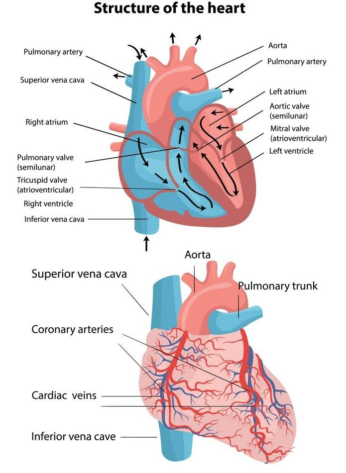
Describe the structure of the Heart with a diagram.
Answer
535.8k+ views
Hint: Heart is a muscular and important organ or major organ of the body. It comes under the circulatory system as its function is to pump blood throughout the body. If a heart stops working the person could die. It is an essential organ. The heart is about the size of our fist located behind and slightly left of the breastbone. Functions of the heart includes pumping oxygenated blood to the other body parts.
Complete answer:
Heart of human beings having four chambered is having two auricles and two ventricles. Heart is a complex organ to discuss about, as it carries all the blood pumps in and is sent out to other body parts. The blood is being carried in closed vessels called capillaries. There are two types of blood capillaries present in the body, one is vein and the other is artery.
Sino-auricular node generates the nerve impulses which are responsible for starting the contraction of the heart. It controls the rhythm of the heart contraction.
Purkinje’s fibers are specialized conducting fibers composed of electrically fitted fibers composed of electrically excited cells. They are myofibrils with mitochondria which conduct cardiac action potentials more quickly and efficiently than any other cells in the heart.
Note:
The walls of the heart are made up of three layers- epicardium, myocardium and endocardium. The upper part of the heart is located at the attachment point of several large blood vessels- the vena cava aorta and pulmonary trunk. The heart has four chambers as we know, two upper atria the receiving chambers and two lower ventricles the discharging chambers.
Recently Updated Pages
Master Class 11 Social Science: Engaging Questions & Answers for Success
Master Class 11 English: Engaging Questions & Answers for Success
Master Class 11 Maths: Engaging Questions & Answers for Success
Master Class 11 Chemistry: Engaging Questions & Answers for Success
Master Class 11 Biology: Engaging Questions & Answers for Success
Master Class 11 Physics: Engaging Questions & Answers for Success
Trending doubts
Differentiate between an exothermic and an endothermic class 11 chemistry CBSE
One Metric ton is equal to kg A 10000 B 1000 C 100 class 11 physics CBSE
Difference Between Prokaryotic Cells and Eukaryotic Cells
There are 720 permutations of the digits 1 2 3 4 5 class 11 maths CBSE
Draw a diagram of a plant cell and label at least eight class 11 biology CBSE
Two of the body parts which do not appear in MRI are class 11 biology CBSE