


The size of pupil is controlled by the
A. Ciliary muscles
B. Suspensory ligaments
C. Cornea
D. Iris muscles
Answer
622.2k+ views
Hint: A pupil is the part through which light enters into the eye. It is located at the center of the iris and it can be adjusted according to the amount of light required by the eye to get a clear image of the object.
Complete answer:
An eye is the organ of sight. The basic structure of an eye consists of the ciliary body, cornea, pupil, iris, lens, retina, macula, optic nerve, choroid, vitreous and aqueous humor, fovea centralis, and photoreceptors.
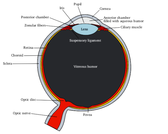
Ciliary muscles are the part of the ciliary body in the eye. It is known to perform the function of changing the shape of the lens when the eye needs to focus on the near object.
Suspensory ligaments are composed of a series of fibers that are known for connecting the ciliary body of the eye with the lens to hold it in its place.
The cornea is the eye’s outermost lens. It is the transparent layer that covers the front portion of the eye. The function of the cornea is to refract or bend the light thereby, focusing most of the light that is entering the eye.
Iris muscles are the two sheets of muscles in the iris with contrary actions i.e., dilation and contraction. These muscles are known for controlling the size of the pupil and thereby determining the amount of light that is reaching the sensory tissue of the retina. Iris is located between the cornea and lens and is the pigmented portion of the eye.
Hence, the correct option is (D).
Note: A pupil’s size is controlled by the two types of iris muscles i.e., radial and circular muscles. Radial muscles are known for contracting in the dim light while the circular muscles are known for contracting in the bright light.
Complete answer:
An eye is the organ of sight. The basic structure of an eye consists of the ciliary body, cornea, pupil, iris, lens, retina, macula, optic nerve, choroid, vitreous and aqueous humor, fovea centralis, and photoreceptors.
Ciliary muscles are the part of the ciliary body in the eye. It is known to perform the function of changing the shape of the lens when the eye needs to focus on the near object.
Suspensory ligaments are composed of a series of fibers that are known for connecting the ciliary body of the eye with the lens to hold it in its place.
The cornea is the eye’s outermost lens. It is the transparent layer that covers the front portion of the eye. The function of the cornea is to refract or bend the light thereby, focusing most of the light that is entering the eye.
Iris muscles are the two sheets of muscles in the iris with contrary actions i.e., dilation and contraction. These muscles are known for controlling the size of the pupil and thereby determining the amount of light that is reaching the sensory tissue of the retina. Iris is located between the cornea and lens and is the pigmented portion of the eye.
Hence, the correct option is (D).
Note: A pupil’s size is controlled by the two types of iris muscles i.e., radial and circular muscles. Radial muscles are known for contracting in the dim light while the circular muscles are known for contracting in the bright light.
Recently Updated Pages
Master Class 12 Business Studies: Engaging Questions & Answers for Success
Master Class 12 Chemistry: Engaging Questions & Answers for Success
Master Class 12 Biology: Engaging Questions & Answers for Success
Class 12 Question and Answer - Your Ultimate Solutions Guide
Master Class 11 English: Engaging Questions & Answers for Success
Master Class 11 Social Science: Engaging Questions & Answers for Success
Trending doubts
Which are the Top 10 Largest Countries of the World?
Draw a labelled sketch of the human eye class 12 physics CBSE
Differentiate between homogeneous and heterogeneous class 12 chemistry CBSE
Differentiate between Pyramid of energy and pyramid class 12 biology CBSE
Why is the cell called the structural and functional class 12 biology CBSE
Draw the diagram of the pyramid of energy Explain In class 12 biology CBSE