


Peyer’s patches are found in
(a) Lamina propria of ileum
(b) Submucosa of ileum
(c) Serosa of jejunum
(d) Muscularis of stomach
Answer
605.4k+ views
Hint: It is organized lymphoid follicles. They are a crucial part of gut-associated lymphatic tissue usually found in humans within the lowest portion of the tiny intestine. It is thick and elongated, found in the intestinal epithelium. It is found that about 100 are found in humans.
Complete answer:
Peyer’s patches are found in the small intestine, it is a secondary lymphoid tissue and it is the component of GALT. They are groups of lymphatic tissues in the submucosa layer of the ileum of the small digestive tract.
Additional information: Peyer's patches are secured by an exceptional follicle-related epithelium that contains particular cells called microfold cells (M cells) which test antigen legitimately from the lumen and convey it to antigen-introducing cells (located during a unique pocket-like structure on their basolateral side) .
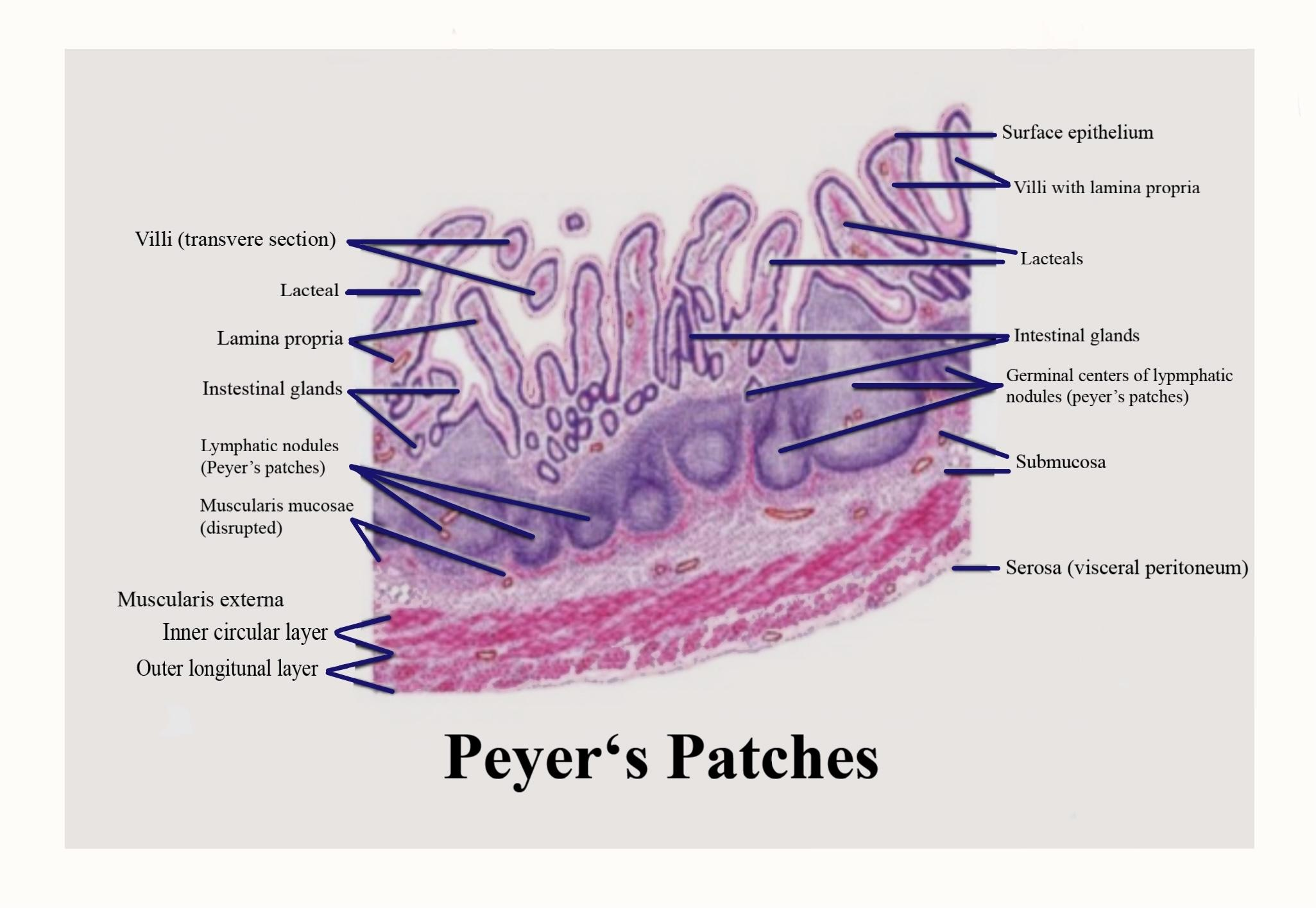
Dendritic cells and macrophages additionally can straightforwardly test the lumen by expanding dendrites through transcellular M cell-explicit pores. At an identical time, the paracellular pathway of follicle-related epithelium is shut firmly to stop the infiltration of antigens and ceaseless contact with immune cells. Immune system microorganisms, B-cells, and memory cells are invigorated after experiencing antigen in Peyer's patches. These cells then pass to the mesenteric lymph nodes where the immune reaction is amplified. Enacted lymphocytes pass into the circulatory system through the lymph vessel and visit the gut where they play out their last effector capacities. The maturation of B-lymphocytes takes place within the Peyer's patch.
So the correct answer is ‘(b) Submucosa of ileum’.
Note: The characteristic feature of the Peyer's patches is the presence of follicle-associated epithelium (FAE), that is found to cover all lymphoid follicles. FAE differs from typical small intestinal villus epithelium: it's fewer goblet cells therefore mucus layer is thinner, and it's also characterized by the presence of specialized M cells or microfold cells, which provide uptake and transport of antigens from the lumen. Besides, basal lamina of follicle-related epithelium is more permeable contrasted with the intestinal villus.
Complete answer:
Peyer’s patches are found in the small intestine, it is a secondary lymphoid tissue and it is the component of GALT. They are groups of lymphatic tissues in the submucosa layer of the ileum of the small digestive tract.
Additional information: Peyer's patches are secured by an exceptional follicle-related epithelium that contains particular cells called microfold cells (M cells) which test antigen legitimately from the lumen and convey it to antigen-introducing cells (located during a unique pocket-like structure on their basolateral side) .
Dendritic cells and macrophages additionally can straightforwardly test the lumen by expanding dendrites through transcellular M cell-explicit pores. At an identical time, the paracellular pathway of follicle-related epithelium is shut firmly to stop the infiltration of antigens and ceaseless contact with immune cells. Immune system microorganisms, B-cells, and memory cells are invigorated after experiencing antigen in Peyer's patches. These cells then pass to the mesenteric lymph nodes where the immune reaction is amplified. Enacted lymphocytes pass into the circulatory system through the lymph vessel and visit the gut where they play out their last effector capacities. The maturation of B-lymphocytes takes place within the Peyer's patch.
So the correct answer is ‘(b) Submucosa of ileum’.
Note: The characteristic feature of the Peyer's patches is the presence of follicle-associated epithelium (FAE), that is found to cover all lymphoid follicles. FAE differs from typical small intestinal villus epithelium: it's fewer goblet cells therefore mucus layer is thinner, and it's also characterized by the presence of specialized M cells or microfold cells, which provide uptake and transport of antigens from the lumen. Besides, basal lamina of follicle-related epithelium is more permeable contrasted with the intestinal villus.
Recently Updated Pages
Master Class 11 English: Engaging Questions & Answers for Success
Master Class 11 Social Science: Engaging Questions & Answers for Success
Master Class 11 Maths: Engaging Questions & Answers for Success
Master Class 11 Biology: Engaging Questions & Answers for Success
Master Class 11 Physics: Engaging Questions & Answers for Success
Master Class 11 Chemistry: Engaging Questions & Answers for Success
Trending doubts
One Metric ton is equal to kg A 10000 B 1000 C 100 class 11 physics CBSE
Difference Between Prokaryotic Cells and Eukaryotic Cells
Find the value of the expression given below sin 30circ class 11 maths CBSE
Difference between physical and chemical change class 11 chemistry CBSE
Two of the body parts which do not appear in MRI are class 11 biology CBSE
Draw a diagram of a plant cell and label at least eight class 11 biology CBSE