




Myofibrils show dark and light bands in
(a) Cardiac muscles
(b) Unstriped muscles
(c) Striped muscles
(d) Both A and C
Answer
 Verified
619.5k+ views
Hint: It runs the whole length of the muscle cell, and since they're only approximately 1.2 µm in diameter, hundreds to thousands are often found inside one muscle cell. They attach to the sarcolemma at their ends, in order that as myofibrils shorten, the whole muscle fiber contracts.
Complete answer:
- The striated appearance of skeletal and heart muscle tissue is a consequence of rehashing groups of the proteins actin and myosin that are available along the length of the myofibril.
-The dark band is known as ‘A’ or Anisotropic band which carries Myosin whereas the light bands which carry actin and are called I-band or Isotropic band. Hence called striped muscles thanks to the presence of striations.
-Hence Myofibrils show dark and lightweight bands in Cardiac muscles and striated muscles.
Additional Information: Myofibrils are contractile units within the cell which contains a daily array of protein myofilaments. Each myofilament runs longitudinally with reference to the muscle cell. There are two types: thick bands and thin bands. Thick bands are made from multiple molecules of a protein called myosin. The thin bands are made from multiple molecules of a protein called actin. The slight actin groups are appended to a Z-line or Z-circle of a versatile protein called titin. From each Z-line to subsequent may be a unit called the sarcomere. The sarcomere is the smallest contractile unit within the myofibril. Sarcomeres contract because the Z-lines move closer together. As the sarcomeres contract the myofibrils contract. As the myofibrils contract the muscle cell contracts. And because the cells contract the whole muscle contracts.
So the correct answer to the above question is ‘Both A and C’.
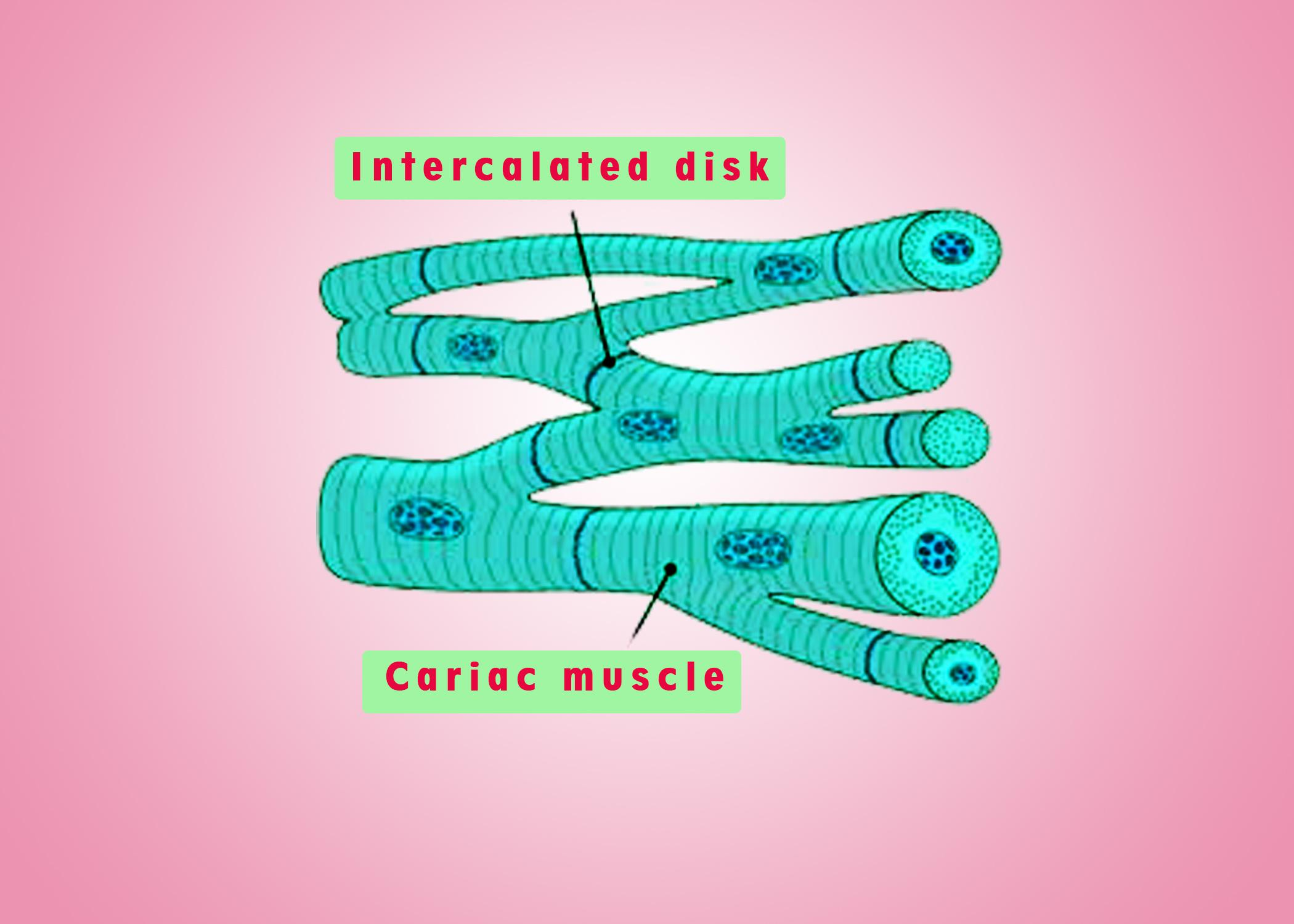
Note: The arrangement of the thick myosin filaments across the myofibrils and therefore the cell causes them to refract light and produce a dark band referred to as the A Band. In between the, A band is a light area where there are no thick myofilaments, only thin actin filaments. These are called the I Bands. The dull groups are the striations seen with the light magnifying lens.
Complete answer:
- The striated appearance of skeletal and heart muscle tissue is a consequence of rehashing groups of the proteins actin and myosin that are available along the length of the myofibril.
-The dark band is known as ‘A’ or Anisotropic band which carries Myosin whereas the light bands which carry actin and are called I-band or Isotropic band. Hence called striped muscles thanks to the presence of striations.
-Hence Myofibrils show dark and lightweight bands in Cardiac muscles and striated muscles.
Additional Information: Myofibrils are contractile units within the cell which contains a daily array of protein myofilaments. Each myofilament runs longitudinally with reference to the muscle cell. There are two types: thick bands and thin bands. Thick bands are made from multiple molecules of a protein called myosin. The thin bands are made from multiple molecules of a protein called actin. The slight actin groups are appended to a Z-line or Z-circle of a versatile protein called titin. From each Z-line to subsequent may be a unit called the sarcomere. The sarcomere is the smallest contractile unit within the myofibril. Sarcomeres contract because the Z-lines move closer together. As the sarcomeres contract the myofibrils contract. As the myofibrils contract the muscle cell contracts. And because the cells contract the whole muscle contracts.
So the correct answer to the above question is ‘Both A and C’.
Note: The arrangement of the thick myosin filaments across the myofibrils and therefore the cell causes them to refract light and produce a dark band referred to as the A Band. In between the, A band is a light area where there are no thick myofilaments, only thin actin filaments. These are called the I Bands. The dull groups are the striations seen with the light magnifying lens.
Recently Updated Pages
Master Class 11 English: Engaging Questions & Answers for Success

Master Class 11 Physics: Engaging Questions & Answers for Success
Master Class 11 Computer Science: Engaging Questions & Answers for Success
Master Class 11 Chemistry: Engaging Questions & Answers for Success
Master Class 11 Social Science: Engaging Questions & Answers for Success
Master Class 11 Accountancy: Engaging Questions & Answers for Success
Trending doubts
Difference Between Prokaryotic Cells and Eukaryotic Cells
Two of the body parts which do not appear in MRI are class 11 biology CBSE
One Metric ton is equal to kg A 10000 B 1000 C 100 class 11 physics CBSE
10 examples of friction in our daily life
Draw a diagram of nephron and explain its structur class 11 biology CBSE
Draw a diagram of a plant cell and label at least eight class 11 biology CBSE