


Formation of rudimentary eyes shows the evolution in an organism is
A. Primary stage of evolution
B. Intermediate stage of evolution
C. Final stage of evolution
D. None of the above
Answer
629.4k+ views
Hint: Our eyes are similar to a tiny camera that receives the light through the sensor called photoreceptors and sends it to the brain to build images.
Complete answer:
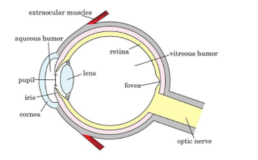
Inside eye following structures are found and these are:
A. A lens
B. Retina
C. Vitreous humor
Evolution of eye-
In evolution in unicellular organisms, we first notice the earliest version of the eye called ‘eyespots’. These are patches of photoreceptor proteins that are photosensitive. Though they don’t help them to see shapes or colour, but helped them to determine the intensity of light. With that help, they could make use of photosynthesis to obtain nutrition.
Over time, these organisms evolved & so do these eyespots also evolved. During the evolution, a depression formed around the eyespot that made the ‘vision’ a little sharper. An opening created in the pit and pupil or the opening through which the light would enter was created. This was followed by the development of the retina and a lens.
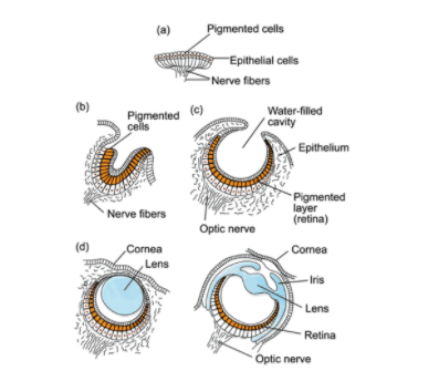
Fig: Stages in the evolution of the eye.
>So, the correct answer is (B) Intermediate stage of evolution
Note: The photoreceptor cell is specialized contains:
a) The opsin, a light-sensitive protein.
b) Chromophore, light-absorbing pigment.
Photon absorbed by the chromophore initiates a chemical reaction that converts the photon's energy into electrical energy and relayed to the nervous system. The photoreceptor cells belong to the retina. The retina is a thin layer of cells that relays visual information to the brain such as the light and day-length information to regulate the circadian rhythm.
In the vertebrates, lenses are composed of epithelial cells that bear the crystallins protein. They are of two types, the α-crystallins and the βγ-crystallins. The crystallins are unique because they are needed for transparency and other lens functions such as tight packing, crystallization resistance, and longevity, so that they must survive entirely in an organism's life. The refractive index gradient is made by the relative distribution of crystallins protein.
Complete answer:
Inside eye following structures are found and these are:
A. A lens
B. Retina
C. Vitreous humor
Evolution of eye-
In evolution in unicellular organisms, we first notice the earliest version of the eye called ‘eyespots’. These are patches of photoreceptor proteins that are photosensitive. Though they don’t help them to see shapes or colour, but helped them to determine the intensity of light. With that help, they could make use of photosynthesis to obtain nutrition.
Over time, these organisms evolved & so do these eyespots also evolved. During the evolution, a depression formed around the eyespot that made the ‘vision’ a little sharper. An opening created in the pit and pupil or the opening through which the light would enter was created. This was followed by the development of the retina and a lens.
Fig: Stages in the evolution of the eye.
>So, the correct answer is (B) Intermediate stage of evolution
Note: The photoreceptor cell is specialized contains:
a) The opsin, a light-sensitive protein.
b) Chromophore, light-absorbing pigment.
Photon absorbed by the chromophore initiates a chemical reaction that converts the photon's energy into electrical energy and relayed to the nervous system. The photoreceptor cells belong to the retina. The retina is a thin layer of cells that relays visual information to the brain such as the light and day-length information to regulate the circadian rhythm.
In the vertebrates, lenses are composed of epithelial cells that bear the crystallins protein. They are of two types, the α-crystallins and the βγ-crystallins. The crystallins are unique because they are needed for transparency and other lens functions such as tight packing, crystallization resistance, and longevity, so that they must survive entirely in an organism's life. The refractive index gradient is made by the relative distribution of crystallins protein.
Recently Updated Pages
Master Class 12 Economics: Engaging Questions & Answers for Success
Master Class 12 English: Engaging Questions & Answers for Success
Master Class 12 Social Science: Engaging Questions & Answers for Success
Master Class 12 Maths: Engaging Questions & Answers for Success
Master Class 12 Physics: Engaging Questions & Answers for Success
Master Class 9 General Knowledge: Engaging Questions & Answers for Success
Trending doubts
Explain the Treaty of Vienna of 1815 class 10 social science CBSE
Why is it 530 pm in india when it is 1200 afternoon class 10 social science CBSE
What is the full form of POSCO class 10 social science CBSE
Define Potential, Developed, Stock and Reserved resources
Which Country Has the Largest Border with India?
Complete the sentence with the most appropriate word class 10 english CBSE