


Draw a well labelled diagram of the human eye and write functions of the following parts: retina, pupil, ciliary muscles and iris.
Answer
570.6k+ views
Hint: Sensory organs are the specialized organs that are composed of sensory neurons that help us to perceive and respond to our surroundings. These organs involve eyes, ears, nose, tongue, and skin.
Complete answer:
Eyes are part of the sensory nervous system that reacts to light and allows vision. There are a pair of eyes located in sockets of the skull called orbits. The movement of eyes assisted by the Six extraocular muscles.
Structure and function of eye parts:
The wall of an eyeball is made up of three layers. The outer wall is relatively tough and white in color called the sclera. The eye consists of various parts:
> Cornea: The anterior part of the sclera is a cornea that serves as a protective covering for the front of the eye and it also helps in focusing light on the retina at the back of the eye.
> Pupil: It is the small aperture in the iris and appears as the black dot in the middle eye. When the amount of light in the immediate surroundings changes, the pupil dilates and constricts like the opening of a camera lens.
> Iris: It is the circular and colored area of the eye which controls the amount of light that enters the eye. When the environment is dark it dilates the pupil to allow more light into the eye and in bright light, it constricts the pupil to enter less light into the eye.
> Ciliary muscles: The choroid layer becomes thick in the anterior part of the cornea that forms the ciliary muscles. These muscles hold the lens in positions.
> Retina: The inner layer of the eye is known as retina and it further consists of three layers- ganglion cells, bipolar cells, and photoreceptor cells from inside to outside. The most sensitive part of this retina is the macula. The retina contains the light-sensing cells that sense the light and blood vessels to nourish these cells.
Note:
- There are two types of photoreceptors named as cones and rods that contain the light-sensitive proteins called the photopigments.
- These photoreceptors convert the image into electrical signals, which are carried to the brain by the optic nerve.
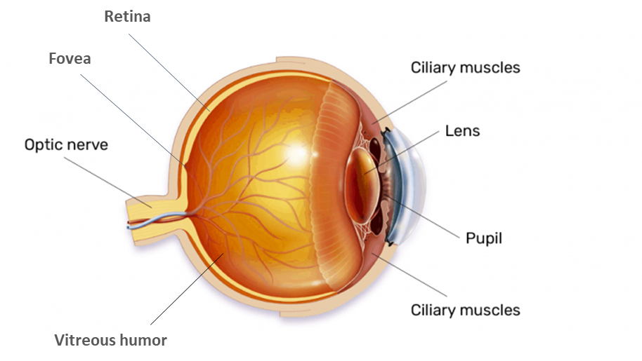
Figure: Structure of a human eye
Complete answer:
Eyes are part of the sensory nervous system that reacts to light and allows vision. There are a pair of eyes located in sockets of the skull called orbits. The movement of eyes assisted by the Six extraocular muscles.
Structure and function of eye parts:
The wall of an eyeball is made up of three layers. The outer wall is relatively tough and white in color called the sclera. The eye consists of various parts:
> Cornea: The anterior part of the sclera is a cornea that serves as a protective covering for the front of the eye and it also helps in focusing light on the retina at the back of the eye.
> Pupil: It is the small aperture in the iris and appears as the black dot in the middle eye. When the amount of light in the immediate surroundings changes, the pupil dilates and constricts like the opening of a camera lens.
> Iris: It is the circular and colored area of the eye which controls the amount of light that enters the eye. When the environment is dark it dilates the pupil to allow more light into the eye and in bright light, it constricts the pupil to enter less light into the eye.
> Ciliary muscles: The choroid layer becomes thick in the anterior part of the cornea that forms the ciliary muscles. These muscles hold the lens in positions.
> Retina: The inner layer of the eye is known as retina and it further consists of three layers- ganglion cells, bipolar cells, and photoreceptor cells from inside to outside. The most sensitive part of this retina is the macula. The retina contains the light-sensing cells that sense the light and blood vessels to nourish these cells.
Note:
- There are two types of photoreceptors named as cones and rods that contain the light-sensitive proteins called the photopigments.
- These photoreceptors convert the image into electrical signals, which are carried to the brain by the optic nerve.
Figure: Structure of a human eye
Recently Updated Pages
Master Class 12 Economics: Engaging Questions & Answers for Success
Master Class 12 Biology: Engaging Questions & Answers for Success
Master Class 11 English: Engaging Questions & Answers for Success
Master Class 11 Physics: Engaging Questions & Answers for Success
Master Class 11 Computer Science: Engaging Questions & Answers for Success
Master Class 11 Chemistry: Engaging Questions & Answers for Success
Trending doubts
What is the Total Duration of Football Match?
Explain the Treaty of Vienna of 1815 class 10 social science CBSE
In football, which nation is called "La Roja"?
Why is there a time difference of about 5 hours between class 10 social science CBSE
10 examples of evaporation in daily life with explanations
What is the full form of POSCO class 10 social science CBSE