


Discuss the internal structure of monocot roots
Answer
627k+ views
Hint: Monocots or monocotyledons are the angiosperms that contain only one cotyledon or embryonic leaf in their seeds. They fundamentally differ from dicots in both morphology and physiology. The monocot roots have a different structure than the dicot roots with visible differentiation in vascular structures too.
Complete answer:
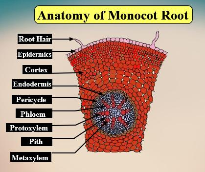
The internal structure of a monocot root consists of the following parts or structures from outside to inside:
-Epiblema/Rhizodermis:
It is the outermost layer of compact parenchymatous cells with no intercellular spaces. The Cuticles and stomata are absent. The root hairs are tubular and unicellular. Epiblema and root hairs help in the absorption of water and minerals.
-Cortex:
The cortex consists of thin-walled rounded or oval-shaped multilayered parenchyma cells with sufficient intercellular spaces. Starch grains are abundantly present in this layer.
Functions of the cortex are: food storage, protection from injuries, conduction of water and minerals to inner tissues.
-Endodermis:
It is the innermost layer of the cortex and is composed of a single layer of barrel-shaped compact cells lacking intercellular spaces.
The radial and the inner tangential walls of endodermal cells are thickened with an internal strip of suberin and lignin. These thickenings are called Casparian strips.
There are some cells opposite to the protoxylem vessels that are not thickened, known as passage cells which serve as a conductive pathway for fluids.
-Stele:
They are the tissues that are present inside the endodermis. It includes the pericycle and vascular elements.
Pericycle:
It is composed of single-layered sclerenchyma cells and gives rise to the lateral roots.
Vascular system:
The vascular system consists of alternating strands of xylem and phloem.
In monocots, the vascular tissues are present in a radial arrangement.
The xylem and the phloem are separated by a layer of sclerenchymatous conjunctive tissue.
Xylem may show exarch or polyarch conditions.
-Conjunctive tissues:
In between the xylem and the phloem bundles, the presence of a many-layered parenchymatous or sclerenchymatous tissue is seen. They help in the storage of food and mechanical support.
-Pith:
It is the large central portion of the root, consisting of thin-walled parenchymatous cells with or without intercellular spaces.
Sometimes the pith may become thick-walled and lignified.
They serve to store food and are abundant reserves of starch grains.
Note: -Passage cells are unique to monocot roots and are absent in the dicots.
-They do not show the presence of cork cambium as seen in dicots.
-Monocot roots have a large number of radially arranged vascular bundles as compared to the small number of exarch vascular bundles in the dicots.
-Pith is well developed in monocot roots whereas they are reduced or sometimes absent in dicot roots.
Complete answer:
The internal structure of a monocot root consists of the following parts or structures from outside to inside:
-Epiblema/Rhizodermis:
It is the outermost layer of compact parenchymatous cells with no intercellular spaces. The Cuticles and stomata are absent. The root hairs are tubular and unicellular. Epiblema and root hairs help in the absorption of water and minerals.
-Cortex:
The cortex consists of thin-walled rounded or oval-shaped multilayered parenchyma cells with sufficient intercellular spaces. Starch grains are abundantly present in this layer.
Functions of the cortex are: food storage, protection from injuries, conduction of water and minerals to inner tissues.
-Endodermis:
It is the innermost layer of the cortex and is composed of a single layer of barrel-shaped compact cells lacking intercellular spaces.
The radial and the inner tangential walls of endodermal cells are thickened with an internal strip of suberin and lignin. These thickenings are called Casparian strips.
There are some cells opposite to the protoxylem vessels that are not thickened, known as passage cells which serve as a conductive pathway for fluids.
-Stele:
They are the tissues that are present inside the endodermis. It includes the pericycle and vascular elements.
Pericycle:
It is composed of single-layered sclerenchyma cells and gives rise to the lateral roots.
Vascular system:
The vascular system consists of alternating strands of xylem and phloem.
In monocots, the vascular tissues are present in a radial arrangement.
The xylem and the phloem are separated by a layer of sclerenchymatous conjunctive tissue.
Xylem may show exarch or polyarch conditions.
-Conjunctive tissues:
In between the xylem and the phloem bundles, the presence of a many-layered parenchymatous or sclerenchymatous tissue is seen. They help in the storage of food and mechanical support.
-Pith:
It is the large central portion of the root, consisting of thin-walled parenchymatous cells with or without intercellular spaces.
Sometimes the pith may become thick-walled and lignified.
They serve to store food and are abundant reserves of starch grains.
Note: -Passage cells are unique to monocot roots and are absent in the dicots.
-They do not show the presence of cork cambium as seen in dicots.
-Monocot roots have a large number of radially arranged vascular bundles as compared to the small number of exarch vascular bundles in the dicots.
-Pith is well developed in monocot roots whereas they are reduced or sometimes absent in dicot roots.
Recently Updated Pages
Master Class 11 English: Engaging Questions & Answers for Success
Master Class 11 Social Science: Engaging Questions & Answers for Success
Master Class 11 Maths: Engaging Questions & Answers for Success
Master Class 11 Biology: Engaging Questions & Answers for Success
Master Class 11 Physics: Engaging Questions & Answers for Success
Master Class 11 Chemistry: Engaging Questions & Answers for Success
Trending doubts
One Metric ton is equal to kg A 10000 B 1000 C 100 class 11 physics CBSE
Difference Between Prokaryotic Cells and Eukaryotic Cells
Find the value of the expression given below sin 30circ class 11 maths CBSE
Difference between physical and chemical change class 11 chemistry CBSE
Two of the body parts which do not appear in MRI are class 11 biology CBSE
Draw a diagram of a plant cell and label at least eight class 11 biology CBSE