


Describe the structure and functions of the various parts of the alimentary canal.
Answer
621.6k+ views
Hint: Human digestive system consists of the organs and the associated digestive glands that help in digestion and when combined they all form the alimentary canal. If not for this system the ingested food cannot be broken down or absorbed to reap its nutrients.
Complete Answer:
Let's begin the explanation by learning about the various parts of the alimentary canal along with their functions.
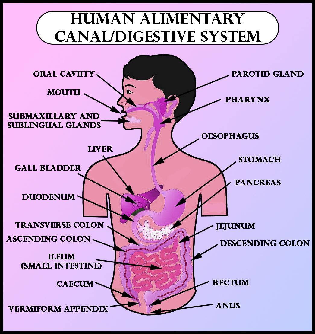
-The alimentary canal begins in the mouth with the buccal cavity or oral cavity, which consists of the teeth and a muscular tongue.
-The hard chewing surface of the teeth, made up of enamel helps in the mastication of the food. The tongue is a muscular organ which is attached to the floor of the mouth with the frenulum.
-Papillae are small projections present on the surface of the tongue on the upper side. Some of these papillae carry taste buds.
-The oral cavity is followed by the pharynx which is a common opening for both oesophagus and trachea. Epiglottis, a cartilaginous flap prevents the entry of food into the glottis during swallowing. -Oesophagus is a thin, long tube which extends from the neck to the thorax and ends in the diaphragm, by opening in the stomach.
-A muscular sphincter called the gastro-oesophageal sphincter regulates the opening and closing of the oesophagus in the stomach.
-The stomach is a j shaped bag like structure located in the upper left portion of the abdomen. It is divided into three regions, namely cardiac, fundic and pyloric.
-The pyloric portion opens in the small intestine, consisting of duodenum, jejunum and ileum. Pyloric sphincters control the opening of the stomach in the small intestine. Ilium opens into the large intestine, which consists of the caecum, colon and rectum.
Now let's look into the microscopic structure of the alimentary canal and their respective functions.
-Right from the oesophagus to rectum, the wall of the alimentary canal possesses four layers namely serosa, muscularis, submucosa and mucosa.
-The outermost layer is ‘serosa’. It is formed from a skinny mesothelium (epithelium of visceral organs) with some connective tissues.
-Muscularis is made by smooth muscles usually arranged into an inner circular and an outer longitudinal layer. An oblique muscle layer could also be present in some regions.
-The submucosal layer is made of loose connective tissues containing nerves, blood and lymph vessels. In the duodenum, glands are also present in submucosa.
-The innermost layer lining the lumen of the alimentary tract is the mucosa. This layer forms irregular folds (rugae) within the stomach and little finger-like foldings called villi within the intestine.
-The cells lining the villi normally produce numerous microscopic projections. These are called 'microvilli.' This arrangement gives a brush border appearance. These modifications increase the surface area enormously for absorption.
-Mucosal epithelium has goblet cells. They secrete mucus that helps in lubrication. Mucosa also forms glands within the stomach (gastric glands) and crypts in between the bases of villi within the intestine (crypts of Lieberkuhn).
Note: -The process of digestion begins in the mouth itself, when the salivary amylase, breaks down starch into maltose.
-Humans have diphyodont, heterodont dentition.
-Liver is the largest gland in the body. It produces bile, which helps in digestion of fat in the duodenum.
-Villi are furnished with a network of capillaries and an outsized lymphatic vessel called the lacteal.
Complete Answer:
Let's begin the explanation by learning about the various parts of the alimentary canal along with their functions.
-The alimentary canal begins in the mouth with the buccal cavity or oral cavity, which consists of the teeth and a muscular tongue.
-The hard chewing surface of the teeth, made up of enamel helps in the mastication of the food. The tongue is a muscular organ which is attached to the floor of the mouth with the frenulum.
-Papillae are small projections present on the surface of the tongue on the upper side. Some of these papillae carry taste buds.
-The oral cavity is followed by the pharynx which is a common opening for both oesophagus and trachea. Epiglottis, a cartilaginous flap prevents the entry of food into the glottis during swallowing. -Oesophagus is a thin, long tube which extends from the neck to the thorax and ends in the diaphragm, by opening in the stomach.
-A muscular sphincter called the gastro-oesophageal sphincter regulates the opening and closing of the oesophagus in the stomach.
-The stomach is a j shaped bag like structure located in the upper left portion of the abdomen. It is divided into three regions, namely cardiac, fundic and pyloric.
-The pyloric portion opens in the small intestine, consisting of duodenum, jejunum and ileum. Pyloric sphincters control the opening of the stomach in the small intestine. Ilium opens into the large intestine, which consists of the caecum, colon and rectum.
Now let's look into the microscopic structure of the alimentary canal and their respective functions.
-Right from the oesophagus to rectum, the wall of the alimentary canal possesses four layers namely serosa, muscularis, submucosa and mucosa.
-The outermost layer is ‘serosa’. It is formed from a skinny mesothelium (epithelium of visceral organs) with some connective tissues.
-Muscularis is made by smooth muscles usually arranged into an inner circular and an outer longitudinal layer. An oblique muscle layer could also be present in some regions.
-The submucosal layer is made of loose connective tissues containing nerves, blood and lymph vessels. In the duodenum, glands are also present in submucosa.
-The innermost layer lining the lumen of the alimentary tract is the mucosa. This layer forms irregular folds (rugae) within the stomach and little finger-like foldings called villi within the intestine.
-The cells lining the villi normally produce numerous microscopic projections. These are called 'microvilli.' This arrangement gives a brush border appearance. These modifications increase the surface area enormously for absorption.
-Mucosal epithelium has goblet cells. They secrete mucus that helps in lubrication. Mucosa also forms glands within the stomach (gastric glands) and crypts in between the bases of villi within the intestine (crypts of Lieberkuhn).
Note: -The process of digestion begins in the mouth itself, when the salivary amylase, breaks down starch into maltose.
-Humans have diphyodont, heterodont dentition.
-Liver is the largest gland in the body. It produces bile, which helps in digestion of fat in the duodenum.
-Villi are furnished with a network of capillaries and an outsized lymphatic vessel called the lacteal.
Recently Updated Pages
Master Class 12 Economics: Engaging Questions & Answers for Success
Master Class 12 English: Engaging Questions & Answers for Success
Master Class 12 Social Science: Engaging Questions & Answers for Success
Master Class 12 Maths: Engaging Questions & Answers for Success
Master Class 12 Physics: Engaging Questions & Answers for Success
Master Class 9 General Knowledge: Engaging Questions & Answers for Success
Trending doubts
Which Country Has the Largest Border with India?
Complete the sentence with the most appropriate word class 10 english CBSE
What were the majoritarian measures taken in Sri Lanka class 10 social science CBSE
The term Eelam means A Country B State C Nation D class 10 social science CBSE
Identify the feminine form of the noun Bachelor a Bachelorette class 10 english CBSE
Identify the feminine form of the word Duke a Dukes class 10 english CBSE