


The T-wave in an ECG represents
A) Electrical excitation of atria
B) Return of the ventricles from excited state
C) Depolarization of ventricles.
D) Beginning of systole
Answer
556.8k+ views
Hint: An ECG is an electrical signal from the heart to check the different heart conditions. ECG stands for electrocardiogram which describes the heart conditions, whether it functions well or not. Electrodes are placed on the chest of the human body to record the heart’s electrical signal, which causes the heart to beat. The machine that records the heart rate or beat of the person is called an electrocardiograph.
Complete answer:
Option A) Electrical excitation of atria.
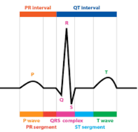
The waves on electrocardiogram include four types of waves- P wave, Q wave, R wave, S wave, T wave and U wave. The P wave represents the electrical excitation of atria or depolarization, which leads to contraction of both atria. As we know how important electrical signals are to the heart. The electrical signal travels through the network of conducting cell pathways which stimulate the upper and lower chambers of the heart which are atria and ventricles respectively. The AV node sends impulse into ventricles and ventricles contract or pump.
So, option A is not correct
Option B) Return of the ventricles from excited state- as we know that heart activity cannot go on a straight line. So, the return of the ventricles from the excited state represents the T wave on the electrocardiogram. It represents the repolarization which returns ventricular myocardium from its excited state. This state denotes when the heart returns back to normal state and starts preparing for contraction again. The T wave has a positive deflection since the cells in the ventricles that depolarize are the first to repolarize.
So, option B is correct.
Option C) Depolarization of ventricles- Depolarization of ventricles are denoted by a series of three deflections that reflect in the right and left ventricles. It denotes the QRS complex or wave in the electrocardiogram. The first deflection in the complex is known as Q wave as its negative. The first positive deflection in the complex is called the R wave. Then, a negative deflection after an R wave is called an S wave. And if there is another positive deflection after the S wave, we call it the R' wave.
So, option c is not correct.
Option D) Beginning of systole- Isovolumetric ventricular contraction marks the beginning of systole and starts with the appearance of the QRS complex. Beginning of systole marks the ventricles to fill during ventricular diastole. After tracing the P wave of the ECG, atria begin to contract. That is known as systole, pulsing blood into ventricles.
There are two phases in systole,
a) Atrial systole which lasts \[0.1\] second, both atria contract and force blood back to ventricles.
b) Ventricular systole- lasts about \[0.3\] seconds, both ventricles contract, blood is forced to the lungs via the pulmonary trunk and rest of the body by aorta.
So, option D is not correct.
So the correct answer is option B i.e., return of the ventricles from excited state.
Note:
The waves of ECG have some time intervals which are denoted by QT and PR intervals. The PR interval runs from the start of the P wave to the start of the QRS. The QT interval is the distance between the start of the QRS and the end of the T wave. It indicates the point at which the ventricles depolarize and repolarize. It is a ventricular action potential measurement (AP).
Complete answer:
Option A) Electrical excitation of atria.
The waves on electrocardiogram include four types of waves- P wave, Q wave, R wave, S wave, T wave and U wave. The P wave represents the electrical excitation of atria or depolarization, which leads to contraction of both atria. As we know how important electrical signals are to the heart. The electrical signal travels through the network of conducting cell pathways which stimulate the upper and lower chambers of the heart which are atria and ventricles respectively. The AV node sends impulse into ventricles and ventricles contract or pump.
So, option A is not correct
Option B) Return of the ventricles from excited state- as we know that heart activity cannot go on a straight line. So, the return of the ventricles from the excited state represents the T wave on the electrocardiogram. It represents the repolarization which returns ventricular myocardium from its excited state. This state denotes when the heart returns back to normal state and starts preparing for contraction again. The T wave has a positive deflection since the cells in the ventricles that depolarize are the first to repolarize.
So, option B is correct.
Option C) Depolarization of ventricles- Depolarization of ventricles are denoted by a series of three deflections that reflect in the right and left ventricles. It denotes the QRS complex or wave in the electrocardiogram. The first deflection in the complex is known as Q wave as its negative. The first positive deflection in the complex is called the R wave. Then, a negative deflection after an R wave is called an S wave. And if there is another positive deflection after the S wave, we call it the R' wave.
So, option c is not correct.
Option D) Beginning of systole- Isovolumetric ventricular contraction marks the beginning of systole and starts with the appearance of the QRS complex. Beginning of systole marks the ventricles to fill during ventricular diastole. After tracing the P wave of the ECG, atria begin to contract. That is known as systole, pulsing blood into ventricles.
There are two phases in systole,
a) Atrial systole which lasts \[0.1\] second, both atria contract and force blood back to ventricles.
b) Ventricular systole- lasts about \[0.3\] seconds, both ventricles contract, blood is forced to the lungs via the pulmonary trunk and rest of the body by aorta.
So, option D is not correct.
So the correct answer is option B i.e., return of the ventricles from excited state.
Note:
The waves of ECG have some time intervals which are denoted by QT and PR intervals. The PR interval runs from the start of the P wave to the start of the QRS. The QT interval is the distance between the start of the QRS and the end of the T wave. It indicates the point at which the ventricles depolarize and repolarize. It is a ventricular action potential measurement (AP).
Recently Updated Pages
Master Class 12 Economics: Engaging Questions & Answers for Success
Master Class 12 English: Engaging Questions & Answers for Success
Master Class 12 Social Science: Engaging Questions & Answers for Success
Master Class 12 Maths: Engaging Questions & Answers for Success
Master Class 12 Physics: Engaging Questions & Answers for Success
Master Class 9 General Knowledge: Engaging Questions & Answers for Success
Trending doubts
One Metric ton is equal to kg A 10000 B 1000 C 100 class 11 physics CBSE
Difference Between Prokaryotic Cells and Eukaryotic Cells
Find the value of the expression given below sin 30circ class 11 maths CBSE
Two of the body parts which do not appear in MRI are class 11 biology CBSE
1 ton equals to A 100 kg B 1000 kg C 10 kg D 10000 class 11 physics CBSE
Draw a diagram of nephron and explain its structur class 11 biology CBSE