


Sinus venosus occurs on
(a) Dorsal side of the heart
(b) Ventral side of the heart
(c) Basal region of the heart
(d) Apical end of heart
Answer
618k+ views
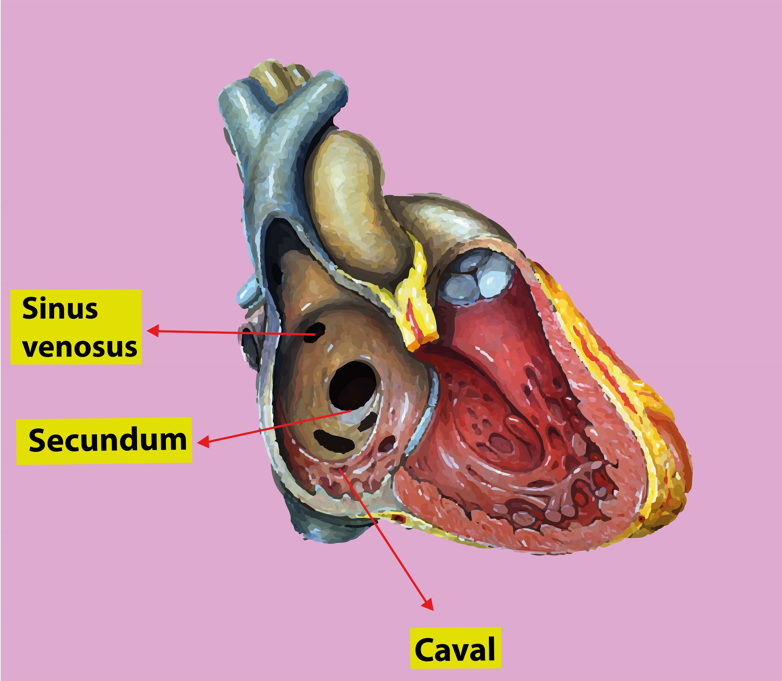
Hint: The sinus venosus is the most caudal of the primitive heart chambers. In humans the fourth week of development, it receives blood from the three sets of veins. The sinus venosus is in continuity with the primitive common atrium.
Complete step by step answer:
Sinus venosus and atrial chamber are at first open communication. They become partially separated by grooves at the junction of these two chambers. Right groove remains shallow, the left one becomes very sharp. Left part becomes completely separated from the atrial chamber. It's right and left horns are prolongations of sinus venosus. The left horn and its tributaries are much reduced in size and appear as tributaries of the right half such that each horn is joined by one vitelline vein from the yolk sac, one vena umbilicalis from the placenta and one common vein from the body wall.
Additional Information: Development of the Sinus Venosus
- In the center of the fourth week, the sinus venosus receives blood from the proper and left sinus horns.
- Each horn receives blood from three important veins: the vitelline or omphalomesenteric vein, the vena umbilicalis and the common vein.
- At first communication between the sinus and therefore the atrium is wide. Soon, however, the doorway of the sinus shifts to the proper.
- This shift is caused primarily by left- to- right shunts of blood, which occur within the venous system during the fourth and fifth weeks of development.
- With the obliteration of the proper vena umbilicalis and therefore the left vitelline vein during the fifth week, the left sinus horn rapidly loses its importance.
- When the left common vein is obliterated at 10 weeks, all that is still of the left sinus horn is that the oblique vein of the left atrium of the heart and therefore the sinus coronarius.
- As a result of left- to- right shunts of blood, the proper sinus horn and veins enlarge greatly.
- The right horn, which now forms the sole communication between the first sinus venosus and therefore the atrium, is incorporated into the proper atrium to make the smooth- walled a part of the proper atrium.
- Its entrance, the sinoatrial orifice, is flanked on all sides by a valvular fold, the proper and left venous valves.
- Dorso Kranial the valves fuse, forming a ridge referred to as the septum spurium.
- Initially, the valves are large, but when the proper sinus horn is incorporated into the wall of the atrium, the left venous valve and therefore the septum spurium fuse with the developing atrial septum.
- The superior portion of the proper venous valve disappears entirely.
- The inferior portion develops into two parts: the valve of the inferior vein and the valve of the sinus coronarius.
- The crista terminalis forms the line between the first trabeculated part of the proper atrium and therefore the smooth- walled part (sinus venarum) , which originates from the proper sinus horn.
So, the correct answer is ‘Dorsal side of the heart.’
Note: The fate of Sinus venosus:
- The right horn forms the graceful posterior wall of the proper atrium.
- The left horn and therefore the body of the sinus venosus atrophy and form the sinus coronarius.
- The left common cardinal veins form the oblique vein of the left atrium of the heart.
Recently Updated Pages
Master Class 12 Economics: Engaging Questions & Answers for Success
Master Class 12 English: Engaging Questions & Answers for Success
Master Class 12 Social Science: Engaging Questions & Answers for Success
Master Class 12 Maths: Engaging Questions & Answers for Success
Master Class 12 Physics: Engaging Questions & Answers for Success
Master Class 11 Social Science: Engaging Questions & Answers for Success
Trending doubts
One Metric ton is equal to kg A 10000 B 1000 C 100 class 11 physics CBSE
Difference Between Prokaryotic Cells and Eukaryotic Cells
Find the value of the expression given below sin 30circ class 11 maths CBSE
1 ton equals to A 100 kg B 1000 kg C 10 kg D 10000 class 11 physics CBSE
Two of the body parts which do not appear in MRI are class 11 biology CBSE
Draw a diagram of nephron and explain its structur class 11 biology CBSE