


Mention the location and function of the given parts:
a) Fovea centralis
b) Choroid
Answer
552.3k+ views
Hint: Fovea centralis and choroid are the important regions that are present in our eye. The Fovea is the area within the retina of the eye where more cone cells are connected and the choroid is the middle layer of the eye which has pigment, vascular and connective tissue.
Complete answer:
To answer this question you must know about the structure of an eye. The human eye is made up of various parts which include Iris, pupil, cornea, lens, choroid, ciliary body, retina, macula, fovea, optic disc, optic nerves, sclera, rod cells and cone cells.
FOVEA CENTRALIS: -
Location (fovea): - it is small tiny parts of eye anatomy located at the centre of the macula and is the area with the great concentration of cone cells.
Function: -the fovea is responsible for sharp vision which is necessary for humans to do various activities such as reading, driving etc.
CHOROID: -
Location (choroid coat):- A thin layer of tissue that is the part of the middle layer of the wall of the eye between the retina and sclera .and it is filled with blood vessels that bring oxygen and nutrients to the eye.
Function: -it contains pigments that help to absorb excess light so it prevents the blurring of vision and nourishes the retina and also maintains the temperature and volume of the eye.
The diagram below shows the structure of the human eye.
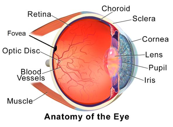
Note: The eye is a complex structure with many small organs that are essential for vision and movements of the eyes. More than 50% of the sensory receptors in the human body are located in the eyes, eyes absorb the visible light from 400 -700 nm. The eye is the source of injury, infection and multiple medical diseases.
Complete answer:
To answer this question you must know about the structure of an eye. The human eye is made up of various parts which include Iris, pupil, cornea, lens, choroid, ciliary body, retina, macula, fovea, optic disc, optic nerves, sclera, rod cells and cone cells.
FOVEA CENTRALIS: -
Location (fovea): - it is small tiny parts of eye anatomy located at the centre of the macula and is the area with the great concentration of cone cells.
Function: -the fovea is responsible for sharp vision which is necessary for humans to do various activities such as reading, driving etc.
CHOROID: -
Location (choroid coat):- A thin layer of tissue that is the part of the middle layer of the wall of the eye between the retina and sclera .and it is filled with blood vessels that bring oxygen and nutrients to the eye.
Function: -it contains pigments that help to absorb excess light so it prevents the blurring of vision and nourishes the retina and also maintains the temperature and volume of the eye.
The diagram below shows the structure of the human eye.
Note: The eye is a complex structure with many small organs that are essential for vision and movements of the eyes. More than 50% of the sensory receptors in the human body are located in the eyes, eyes absorb the visible light from 400 -700 nm. The eye is the source of injury, infection and multiple medical diseases.
Recently Updated Pages
Master Class 5 English: Engaging Questions & Answers for Success
Master Class 5 Maths: Engaging Questions & Answers for Success
Master Class 5 Social Science: Engaging Questions & Answers for Success
Master Class 5 Science: Engaging Questions & Answers for Success
Class 5 Question and Answer - Your Ultimate Solutions Guide
Master Class 9 General Knowledge: Engaging Questions & Answers for Success
Trending doubts
One Metric ton is equal to kg A 10000 B 1000 C 100 class 11 physics CBSE
What is cell theory Who formulated it class 11 biology CBSE
Phyllotaxy is the arrangement of ALeaflets BLeaves class 11 biology CBSE
Difference Between Prokaryotic Cells and Eukaryotic Cells
The symbiotic association of fungi and algae is called class 11 biology CBSE
Cell theory was formulated by A Schleiden and Schwann class 11 biology CBSE