


Give the scientific names of the following parts of the eye:
(a) carries signals from the eye to the brain.
(b) muscles that change the shape of the eye-lens.
(c) a hole in the middle of the iris.
(d) a clear window at the front of the eye.
(e) changes shape to focus a picture on the retina.
Answer
523.8k+ views
Hint: The visual system includes the eyes. They give animals vision, the ability to receive and process visual information, and the ability to perform several photo response functions that are not dependent on vision. Light is detected by the eyes and converted into electrochemical impulses in the neurons.
Complete answer:
Our visual organ is the eye. The cornea, iris, pupil, lens, retina, macula, optic nerve, choroid, and vitreous are just a few of the components that make up the eye.
Carries signals from an eye to the brain: Optic nerve: The second cranial nerve, the optic nerve, carries sensory nerve impulses from the retina's over one million ganglion cells to the visual centers in the brain. The vast majority of optic nerve fibers transmit central vision information.
Muscles that change the shape of the eye-lens: Ciliary muscles: The ciliary muscle is an intrinsic eye muscle that forms a ring of smooth muscle in the middle layer of the eye (vascular layer). It regulates the flow of aqueous humor into Schlemm's canal and controls accommodation for viewing objects at different distances.
A hole in the middle of the iris: Pupil: The pupil is a black hole in the center of the iris that allows light to enter the eye and strike the retina. Light rays entering the pupil are either absorbed directly by the tissues inside the eye or absorbed after diffuse reflections within the eye that mostly miss exiting the narrow pupil, giving it a black appearance.
A clear window at the front of the eye: Cornea: The cornea is the clear, protective outer layer of your eye. It acts as a barrier against dirt, germs, and other potentially harmful substances, working in tandem with the sclera (the white of your eye). Fun fact: Your cornea can also block some ultraviolet light from the sun.
Changes shape to focus a picture on the retina: Eye lens: The lens is made up of transparent, flexible tissue and is situated behind the iris and pupil. After the cornea, it is the second part of your eye that helps focus light and images on your retina.
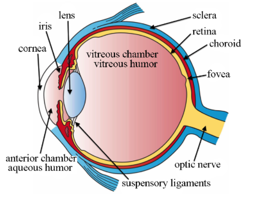
Note: Pit eyes are the most basic eyes. They are eyespots that are placed in a pit to reduce the angles of light that enter and affect the eyespot, allowing the organism to determine the angle of incoming light. Retinal photosensitive ganglion cells send signals to the suprachiasmatic nuclei and the pretectal area via the retinohypothalamic tract in more complex eyes to adjust the circadian adjustment.
Complete answer:
Our visual organ is the eye. The cornea, iris, pupil, lens, retina, macula, optic nerve, choroid, and vitreous are just a few of the components that make up the eye.
Carries signals from an eye to the brain: Optic nerve: The second cranial nerve, the optic nerve, carries sensory nerve impulses from the retina's over one million ganglion cells to the visual centers in the brain. The vast majority of optic nerve fibers transmit central vision information.
Muscles that change the shape of the eye-lens: Ciliary muscles: The ciliary muscle is an intrinsic eye muscle that forms a ring of smooth muscle in the middle layer of the eye (vascular layer). It regulates the flow of aqueous humor into Schlemm's canal and controls accommodation for viewing objects at different distances.
A hole in the middle of the iris: Pupil: The pupil is a black hole in the center of the iris that allows light to enter the eye and strike the retina. Light rays entering the pupil are either absorbed directly by the tissues inside the eye or absorbed after diffuse reflections within the eye that mostly miss exiting the narrow pupil, giving it a black appearance.
A clear window at the front of the eye: Cornea: The cornea is the clear, protective outer layer of your eye. It acts as a barrier against dirt, germs, and other potentially harmful substances, working in tandem with the sclera (the white of your eye). Fun fact: Your cornea can also block some ultraviolet light from the sun.
Changes shape to focus a picture on the retina: Eye lens: The lens is made up of transparent, flexible tissue and is situated behind the iris and pupil. After the cornea, it is the second part of your eye that helps focus light and images on your retina.
Note: Pit eyes are the most basic eyes. They are eyespots that are placed in a pit to reduce the angles of light that enter and affect the eyespot, allowing the organism to determine the angle of incoming light. Retinal photosensitive ganglion cells send signals to the suprachiasmatic nuclei and the pretectal area via the retinohypothalamic tract in more complex eyes to adjust the circadian adjustment.
Recently Updated Pages
Master Class 11 Social Science: Engaging Questions & Answers for Success
Master Class 11 Physics: Engaging Questions & Answers for Success
Master Class 11 Maths: Engaging Questions & Answers for Success
Master Class 11 Economics: Engaging Questions & Answers for Success
Master Class 11 Computer Science: Engaging Questions & Answers for Success
Master Class 11 Chemistry: Engaging Questions & Answers for Success
Trending doubts
One Metric ton is equal to kg A 10000 B 1000 C 100 class 11 physics CBSE
There are 720 permutations of the digits 1 2 3 4 5 class 11 maths CBSE
1 Quintal is equal to a 110 kg b 10 kg c 100kg d 1000 class 11 physics CBSE
State and prove Bernoullis theorem class 11 physics CBSE
Difference Between Prokaryotic Cells and Eukaryotic Cells
Discuss the various forms of bacteria class 11 biology CBSE