


Draw a labelled diagram of the cardiac muscles.
Answer
603k+ views
Hint: In most animals, the heart is a muscular organ that helps in pumping the blood in the circulatory system of animals. The arteries carry the oxygenated blood from the heart to other organs. On the other hand, the veins carry the deoxygenated blood from other organs to the hearts.
Complete answer:
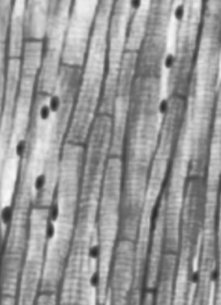
The cardiac muscles are branched and striated. These muscles are uninucleated, that is only a single cell is present inside the. These cells are present inside the heart, thus they are often termed as myocardium or the heart muscle. And they are involuntary in nature.
A thick middle layer is formed by the myocardium, between epicardium (outer layer of the heart) and endocardium (inner layer of the heart). The contraction of cardiac muscles is similar to the skeletal muscles, but there are some differences. In the sarcoplasmic reticulum, the action potential triggers the release of calcium ions with the help of electric stimulation. Excitation contraction couplings happen when the increase in calcium ions causes the myofilaments of the cells to slide past to each other.
The disease of the heart muscles are angina pectoris, myocardial infarction and cardiomyopathies.
Note: Cardiac muscles striated muscles, involuntary muscles, branched, uninucleate, they form an interconnecting network and also self excitatory. There are three types of muscles namely skeletal, smooth and cardiac muscles. The skeletal muscles are striated, multinucleated and tubular fibers. These muscles are voluntary. The smooth muscles are spindle shaped, and non- striated, they have only one nucleus. These muscles are involuntary.
Complete answer:
The cardiac muscles are branched and striated. These muscles are uninucleated, that is only a single cell is present inside the. These cells are present inside the heart, thus they are often termed as myocardium or the heart muscle. And they are involuntary in nature.
A thick middle layer is formed by the myocardium, between epicardium (outer layer of the heart) and endocardium (inner layer of the heart). The contraction of cardiac muscles is similar to the skeletal muscles, but there are some differences. In the sarcoplasmic reticulum, the action potential triggers the release of calcium ions with the help of electric stimulation. Excitation contraction couplings happen when the increase in calcium ions causes the myofilaments of the cells to slide past to each other.
The disease of the heart muscles are angina pectoris, myocardial infarction and cardiomyopathies.
Note: Cardiac muscles striated muscles, involuntary muscles, branched, uninucleate, they form an interconnecting network and also self excitatory. There are three types of muscles namely skeletal, smooth and cardiac muscles. The skeletal muscles are striated, multinucleated and tubular fibers. These muscles are voluntary. The smooth muscles are spindle shaped, and non- striated, they have only one nucleus. These muscles are involuntary.
Recently Updated Pages
Master Class 12 Economics: Engaging Questions & Answers for Success
Master Class 12 English: Engaging Questions & Answers for Success
Master Class 12 Social Science: Engaging Questions & Answers for Success
Master Class 12 Maths: Engaging Questions & Answers for Success
Master Class 12 Physics: Engaging Questions & Answers for Success
Master Class 12 Business Studies: Engaging Questions & Answers for Success
Trending doubts
Which are the Top 10 Largest Countries of the World?
Draw a labelled sketch of the human eye class 12 physics CBSE
Differentiate between homogeneous and heterogeneous class 12 chemistry CBSE
Why is the cell called the structural and functional class 12 biology CBSE
Draw ray diagrams each showing i myopic eye and ii class 12 physics CBSE
Which is the correct genotypic ratio of mendel dihybrid class 12 biology CBSE